3D Visualization of MR Images
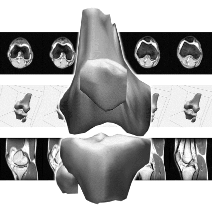
 The femur, tibia, patella, and fibula of my right knee, as reconstructed from the MRI data. Axial and saggittal images from the original tape can be seen in the background.
|
On April 4, 1993, I tore a ligament in my right knee while playing basketball. A few days later I went in for NMR images, and noted that they used 1/2 inch magnetic tape to store them. Trying to turn an unfortunate situation into a chance to do something interesting, I hobbled back to the MRI center with my own tape and asked a gracious but somewhat surprised technician to make a copy of the tape. With the help of System Managers Sharon Taylor and Kevin Mullally, I was able to read the image files from the tape to my home directory. I then deciphered the image format, wrote some interactive feature extraction software, and created a 3D spline model of my knee. In order to create the surface mesh, I designed a relaxation algorithm to form correspondences between the slices. This became my project for Brian Barsky's CS284 geometric modeling class. You're more than welcome to download the dataset of 110 256×256 pixel MR Images in TIF format: debevec_knee_images.tar.gz, 6,803,949 bytes.
 The images are kind of trippy if you play them back as a movie.
|